Opportunity
Brenna Sund (daughter, UNCW intern)
World-renowned heart surgeon, Dr. Randall Wolf, presents ![]() to 35 world-class heart surgeons from such prominent institutions as Cleveland Clinic, Johns Hopkins, DeBakey, University of Chicago, NYU, Rutgers, and Medtronic.
to 35 world-class heart surgeons from such prominent institutions as Cleveland Clinic, Johns Hopkins, DeBakey, University of Chicago, NYU, Rutgers, and Medtronic.
Dr. Wolf’s closing question—with this looping video:
Who would want to visualize flow through graft and coronary for obstructive disease—on the table—after cross-clamp—before coming off pump?
A near unanimous response!
by 21st Century Cardiothoracic Surgical Society | Palm Beach meeting
Jim Sund, Founding CEO/CTO and
Renowned Heart Surgeon Dr. Randall Wolf
New Model
Instructional Demo (original model)
Finally see both velocity & volume flow as vessel volume changes
(ex-vivo porcine heart-coronary)
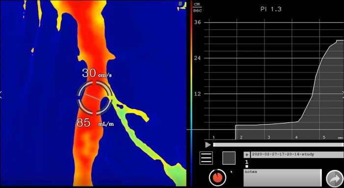
See how blood flows through squeezed tube
See how blood flows around blockage
⇐ (right to left) hemodynamics

Simultaneously see fast, stagnant, and recirculating flow
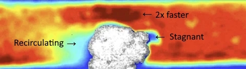
New clinical value: See …
- 2x-3x faster (on the right, upstream side): Incoming flow often accelerates as it approaches a constriction or protrusion, due to conservation of mass (continuity equation)—blood speeds up to maintain volume flow rate through the reduced cross-section. A 2x increase is plausible in simulations of moderate stenoses, where velocities can double or more near the obstacle compared to baseline.
- Recirculating “Eddy currents” (on the left, downstream blue area): This labels the wake region where flow separates, forming eddies or vortices. Blood recirculates in loops, leading to prolonged residence times that promote clotting factors to accumulate.
- Stagnant (near the obstacle on the left): Points to a low-velocity pocket within the recirculation zone, where flow is nearly zero (<5 cm/s or even negative in eddies), exemplifying stasis—one of Virchow’s triad elements for thrombosis.
- Clinical relevance: These are consistent with computational fluid dynamics (CFD) models of pathological blood flow, such as in atherosclerosis or post-stent scenarios, where upstream acceleration contrasts with downstream turbulence and stagnation.
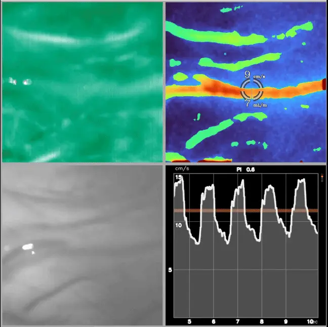
Optional quad view (tissue & vessel)
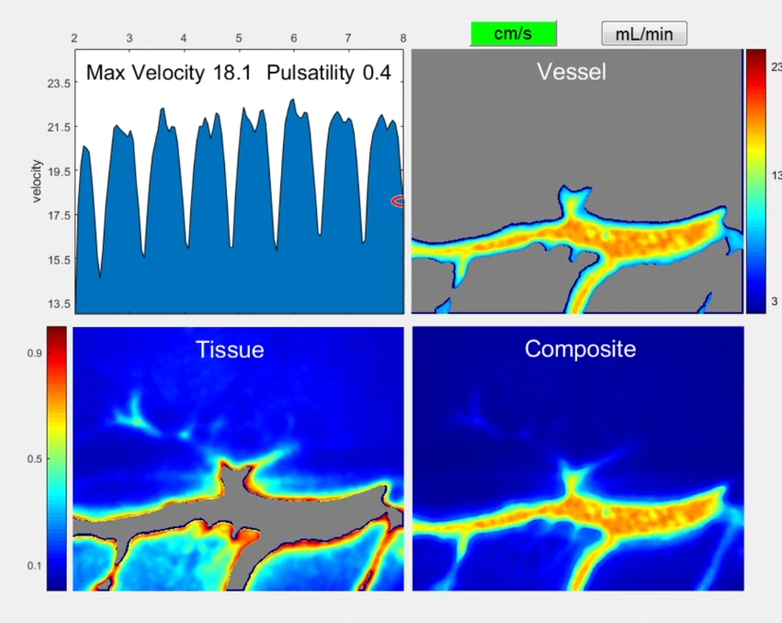
Top left quadrant: Dye doesn’t indicate pulsing flow in artery